Paget’s Disease is a metabolic bone disease most likely caused by a virus and leading to abnormal bone resorption and formation. It is more common in older patients. Paget’s Disease leads to deformities and fractures of bones, as well as arthritis of the hip and knee.
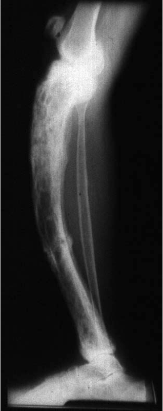
In affected individuals, there is an abnormal breakdown of bone followed by abnormal bone formation. The new bone is larger but weaker and filled with blood vessels. This results in pain, misshapen bones, fractures, and arthritis. In some rare cases, it can develop into bone cancer known as Paget’s Sarcoma.
Paget’s Disease is usually localized, affecting only one or two bones, most commonly the pelvis, femur, humerus, and lower vertebrae. Affected individuals do not display symptoms. The disease is diagnosed when the bone fractures or when an x-ray is taken for another reason.
The mechanism of Paget’s Disease is linked to osteoclasts, which absorb dead bone tissue. The pathogenesis for Paget’s Disease is described in the following steps:
- Increased osteoclasts in specific bone site
- Compensatory increase of osteoblasts
- New bone formation in a “mosaic” pattern
- Breakdown of weak bone resulting in fracture
The disease progression begins with an increase in bone resorption caused by a large accumulation of osteoclasts in a specific bone region. This causes a compensatory increase in osteoblast activity, which form new bone. The new “Pagetic bone” is produced in a mosaic pattern, rather than the normal linear lamellar pattern, and replaces the resorbed bone. The new bone is weaker and susceptible to fractures and arthritis in adjacent joints.