Reconstruction of the hip is called the SUPERhip procedure (SUPER is an acronym for Systematic Utilitarian Procedure for Extremity Reconstruction). Dr. Paley invented the SUPERhip procedure in 1997. It is a combination of surgical procedures designed to comprehensively address and correct severe bone and soft tissue deformities. The SUPERhip can be performed as early as age two and is recommended between the ages of two and three. The SUPERhip can be performed at any age, including adulthood. Getting it done before age three allows one year of recovery before starting the first lengthening. Dr. Paley prefers to start the first lengthening by age 4 whenever possible. The SUPERhip is performed six to twelve months before the first lengthening. To learn more about the SUPERhip, download Dr. Paley's recent book chapter on SUPERhip and SUPERhip2 Procedures for Congenital Femoral Deficiency
SUPER Hip Patient Guide
The SUPERhip procedure has three parts:
- Soft-tissue releases
- Used to correct hip flexion, abduction, and external rotation
- Result: Corrects contractures of hip joint
- Femoral osteotomy
- Used to correct upper femoral varus, flexion, and external rotation
- Result: Corrects bony deformities of the femur
- Pelvic osteotomy
- Used to correct lack of femoral head coverage
- Result: Corrects hip dysplasia
Soft Tissue Releases
Dr. Paley begins by making one long incision from the top of the pelvis to the bottom of the knee. In some cases, if the femur is relatively long, these are broken into two shorter incisions with a space between them. Dr. Paley will then perform the soft-tissue releases, taking great care to avoid harming the femoral and sciatic nerves.
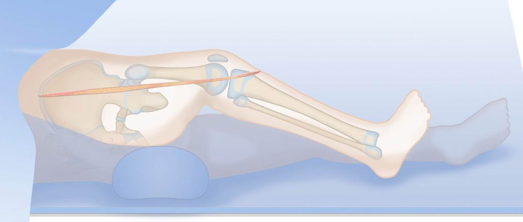
The first soft tissue Dr. Paley identifies is the tensor fascia lata, which is severely contracted. The fascia lata is removed, and if a SUPERknee procedure is required, it is saved for that surgery; if no SUPERknee is necessary, the fascia lata is sutured to the greater trochanter at the end of the procedure.
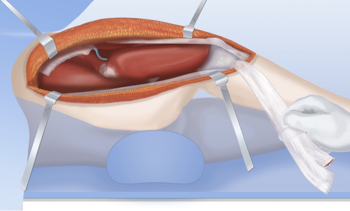
Release of the tensor fascia lata exposes the rectus femoris tendon which is also contracted. The rectus femoris is part of the quadriceps muscle and it is released as well.
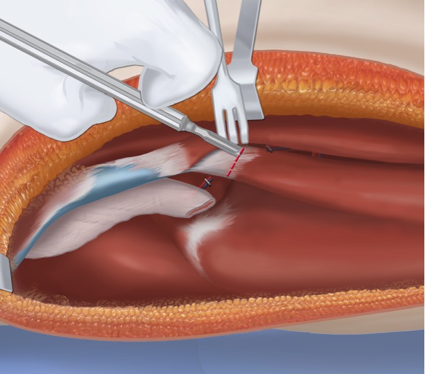
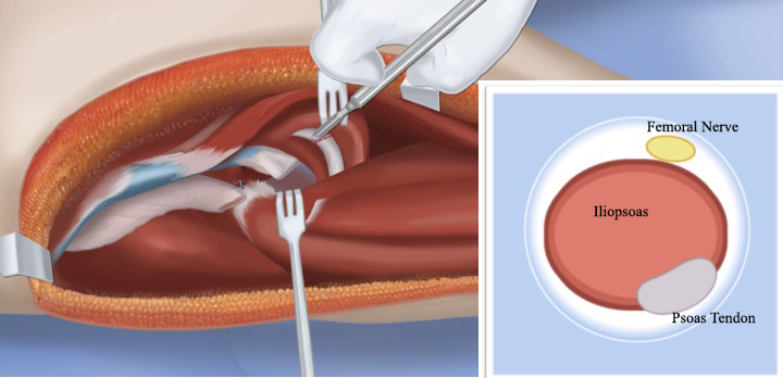
After release of the rectus femoris, Dr. Paley identifies the ilias psoas tendon. The psoas tendon is very close to the femoral nerve. The femoral nerve is carefully moved out of the way and the psoas tendon is cut, leaving the iliacus muscle intact.
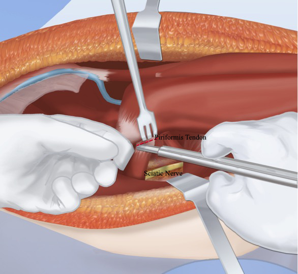
Next, the gluteus maximus muscle is pulled aside to expose the piriformis tendon. The piriformis tendon is what prevents the femur from rotating, resulting in an external rotation contracture. The piriformis tendon is then released and Dr. Paley takes great care to avoid the sciatic nerve.
The next step in the muscle releases is to correct the contracture of the abductor muscles. These muscles are attached to the pelvis. These muscles are very important for hip function. In order to detach the muscles from the pelvis while preserving function and strength, Dr. Paley uses a method he developed called the abductor slide. In the abductor slide, the cartilage at the top of the ilium (pelvic bone) is split and the periosteum and the muscles are peeled back in one motion.
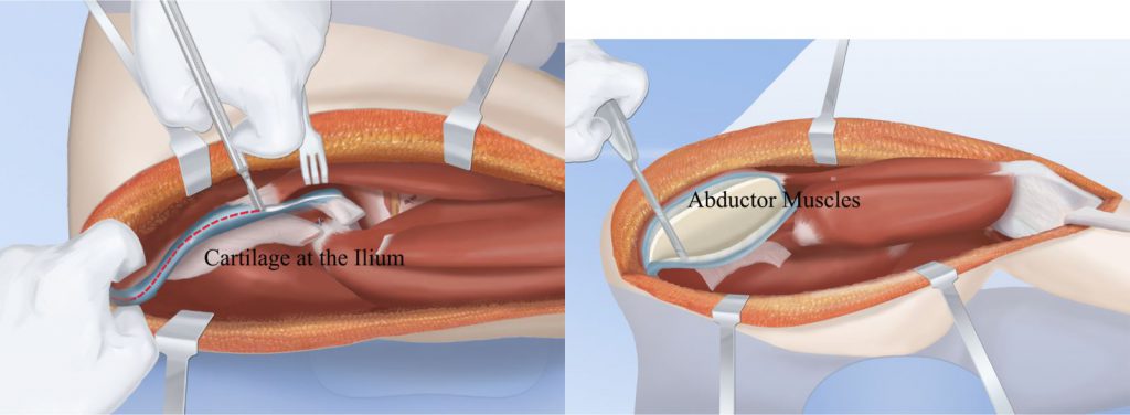
Once these muscles and tendons have been released, the hip can be rotated into the correct anatomic position (out of its deformed position). This completes the first portion of the SUPERhip: soft tissue releases.
Femoral Osteotomy
The second portion of the SUPERhip is the correction of the bony deformity of the upper femur. Patients requiring a SUPERhip have a large bend to the upper femur. This produces a bump that can be felt under the skin. The bump is usually the apex of the deformity and is the location Dr. Paley chooses to perform the osteotomy (surgical cut of the bone). To accurately reposition the upper femur to the exact location of the center of the femoral head, it needs to be visualized during surgery. Rather than surgically opening the hip joint, dye is injected into the joint to visualize the femoral neck and head using a very low-dose x-ray fluoroscopic machine called an image intensifier. This allows accurate placement of hardware into the small femoral head and neck.
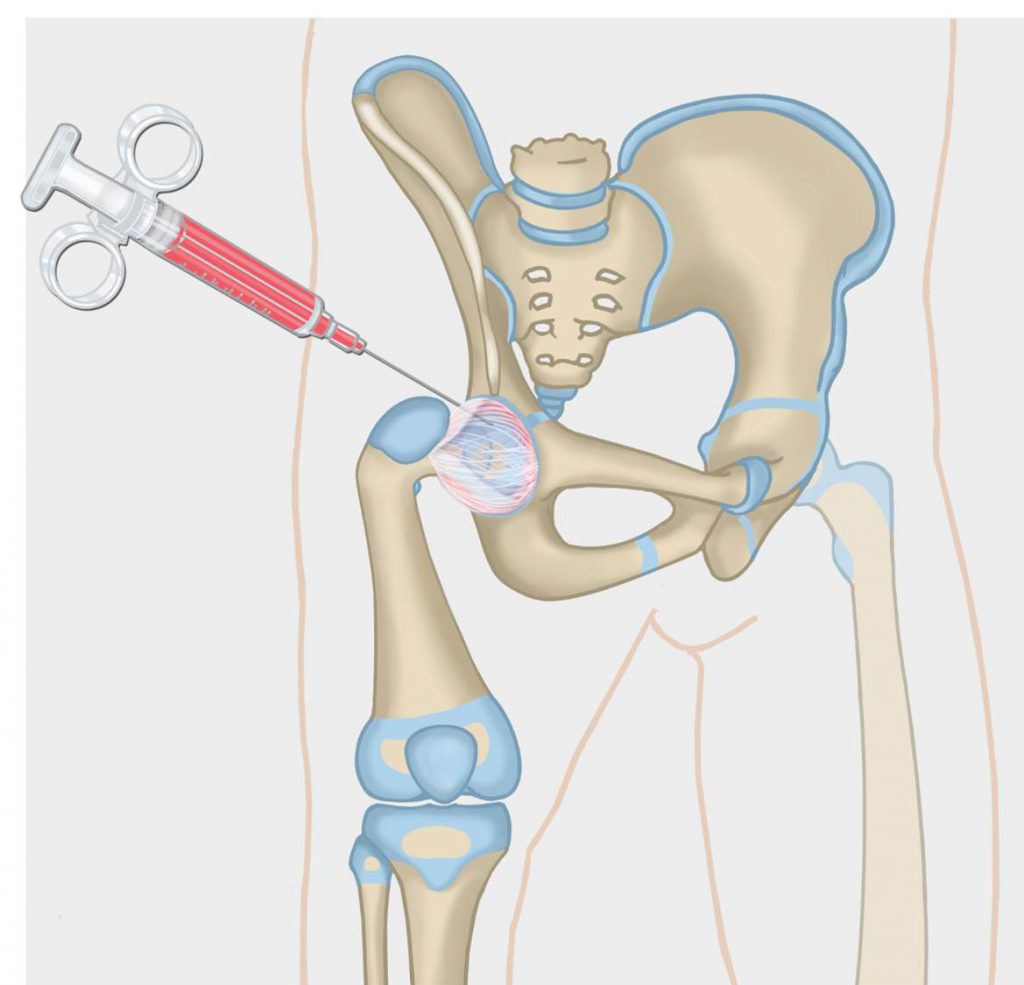
A guide wire is then inserted from the tip of the greater trochanter to the center of the femoral head. A second guide wire is inserted forty-five degrees from the first. This angle is the normal angle between the femoral neck and the shaft of the femur (which is 135 degrees).
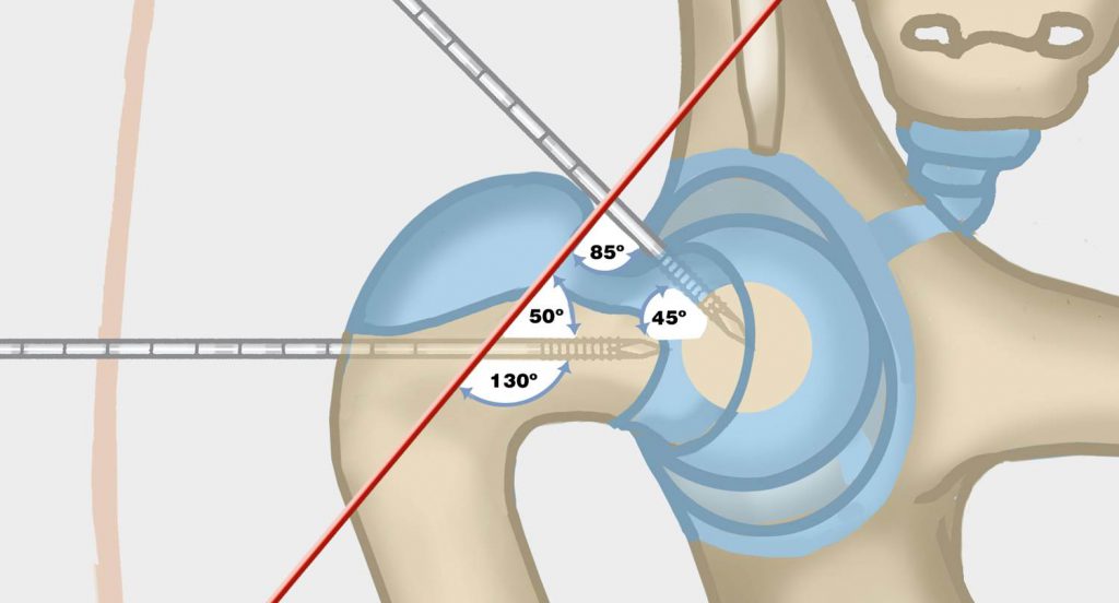
The guide wire at the top of the neck now guides a cannulated chisel that makes a hole in the femoral head through the femoral neck. The chisel is placed perpendicular to the back of the greater trochanter. A cannulated blade plate is then inserted over the guide wire. A wedge of bone is cut parallel and perpendicular to the plate.
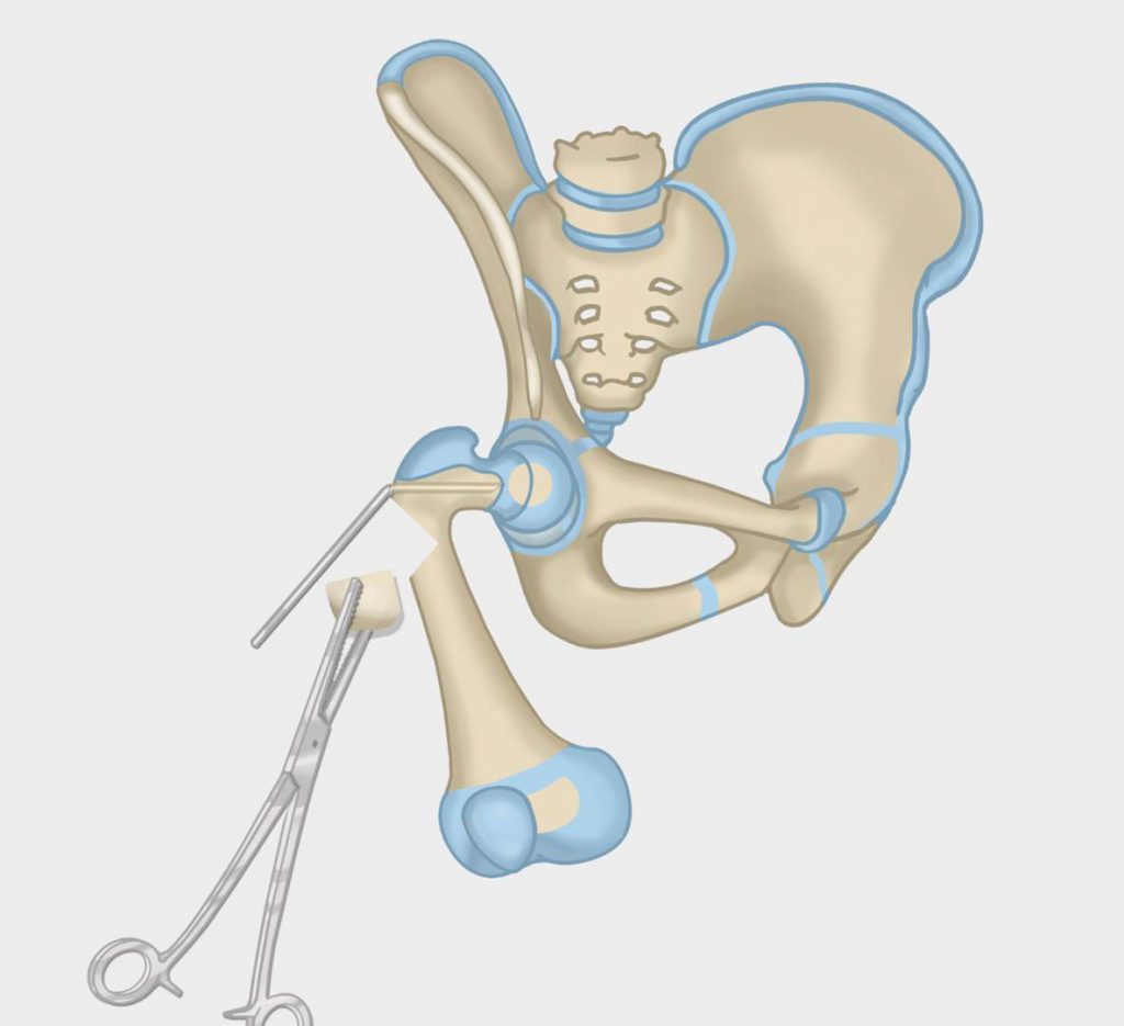
A second osteotomy is then performed obliquely, from the distal edge of the notch across the femur. The femur is then separated and externally rotated and overlapped to the upper portion of the femur. The femur cannot be slotted into place because the muscles, nerves, and arteries are all tethered to the femur.
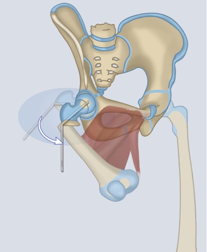
In order to realign the femur, a portion must then be removed. This is done in conjunction with the pelvic osteotomy (described below) and results in reorientation of the neck and head of the femur into its proper position.
Pelvic Osteotomy
The next step is the pelvic osteotomy. In young children, we use Dr. Paley's modification of what is known as the Dega osteotomy. In older children, we use the Periacetabular Triple osteotomy. And in adults and teenagers, we use the Ganz osteotomy.
Dega Osteotomy
The next step is the pelvic osteotomy. In young children, we use Dr. Paley’s modification of what is known as the Dega osteotomy. This is a slanted bone cut that hinges on the growth plate of the acetabulum (hip joint). The cut is circular around the acetabulum, creating a three-dimensional correction and excellent coverage of the femoral head.
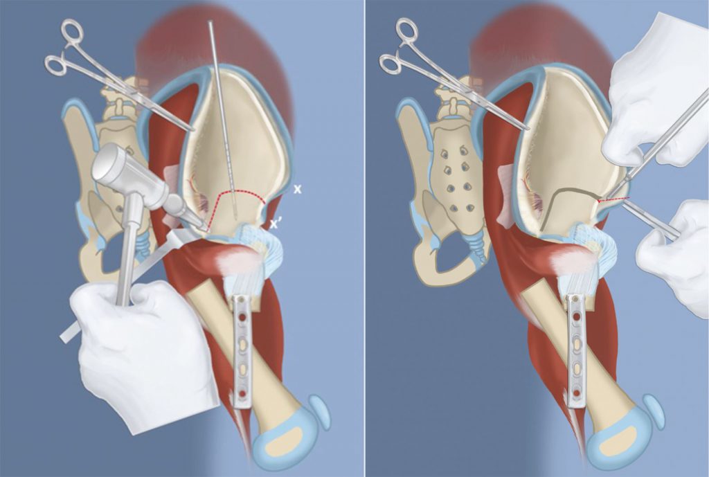
The femur is then oriented into the proper position, overlapping to the upper femur. Since the soft tissues are tethering the femur, it cannot be slotted into place. Instead, a segment of the femur must be removed. This segment of bone will be used to complete the Dega osteotomy. The femur can now be slotted into place.
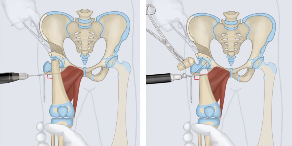
The bone graft from the femur is then inserted into the Dega osteotomy. This restores the normal anatomy of the hip joint and prevents the hip from sliding out of its joint during lengthening.

Next, the femur can be slotted into place. The blade plate now aligns with the femur and screws are inserted through the blade plate into the femur to maintain the alignment. Reorientation actually results in increased length. The femur is the same after as it was before the osteotomy despite the shortening. The reason for this is that the gain in length by reorienting the femoral head and neck is equal to the amount of shortening required to straighten the bone and bring the bone ends together. The femur now looks like a normal femur, albeit short.
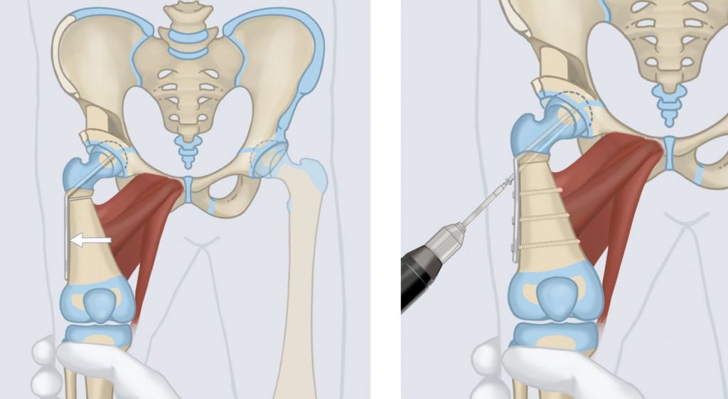
If there is delayed ossification of the femoral neck (Type 1B), the cartilage of the femoral neck needs to be converted into bone. Merely applying the hardware will generally not induce ossification. Lengthening usually cannot proceed until the cartilage of the femoral neck turns into bone (ossifies). In the early days of performing the SUPERhip, this problem lead to recurrence of deformity and breakage of the hardware. One major discovery made by Dr. Paley is that he could get this cartilage to ossify if he inserted bone morphogenic protein (BMP) into the cartilage. BMP is a growth factor that induces bone to grow and is FDA-approved in adult orthopedic surgery to help fractures and nonunions heal or for spine fusion surgery. It is not FDA-approved in children. Doctors are allowed to use it in children as off-label use. Many pediatric orthopedic surgeons use BMP off-label in children for a variety of difficult bone healing problems (e.g. congenital pseudarthrosis of the tibia).
While there are theoretical risks of tumor formation, this has never been seen by pediatric orthopedic surgeons or by Dr. Paley. Dr. Paley’s observation that BMP will cause cartilage to turn into bone was a major discovery that was recently presented at several national and international meetings. To date, Dr. Paley has never had a single complication related to the use of BMP and has used it in over 200 children over the past 10 years. A hole is drilled in the femoral neck and BMP (trade-name INFUSE, made by Medtronics, Memphis, TN) is inserted into this channel via collagen sponges.
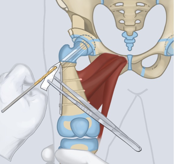
This completes the reconstruction of the hip joint. The final step is to put the muscles back in place. The muscles cannot go back into place because they are tight. To accommodate this, Dr. Paley removes some bone from the top of the iliac crest. This shortens the distance to where the muscles reconnect, in effect loosening the muscles. The iliac bone graft is inserted into the Dega osteotomy site along with the femoral bone graft. This leads to rapid healing of that area.
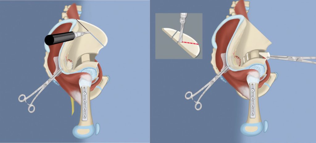
At this point all of the muscles and tendons can be sutured back into place. The apophysis (cartilage of the hip) is sutured back, restoring the proper tension to all the muscles. If the tensor fascia lata is not going to be used for a superknee procedure, it is transferred to the greater trochanter at this time. The incision is then sutured close.
All aspects of the deformities, deficiencies, and instability of the upper femur and hip have now been successfully addressed by the SUPERhip procedure. All that is left is to reconnect the muscles that were released into a better position and tension. This completes the SUPERhip procedure.
Periacetabular Triple Osteotomy
In older children, a different, more complicated osteotomy, called a Paley triple periacetabular osteotomy is performed to gain greater and more three-dimensional reorientation of the acetabulum while sparing the growth plate. In this osteotomy, three separate cuts of the pelvis are performed to release the hip joint.
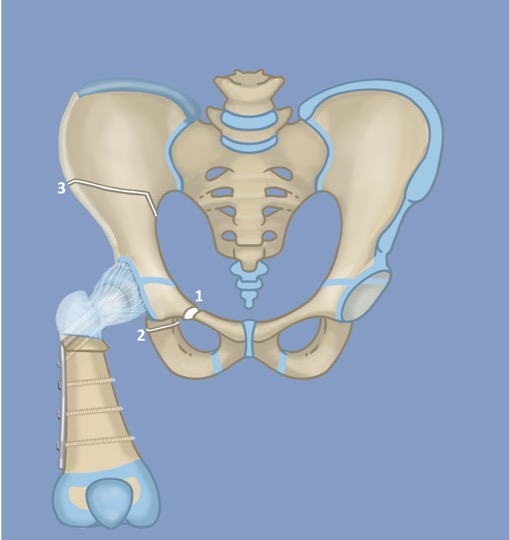
The released hip joint is then rotated twice in two different planes. It helps to visualize the pelvis along three different axes: the X, Y, and Z axes, as demonstrated below:

First, the pelvis is rotated along the Z-axis.

Second, the pelvis is rotated along the Y-axis.
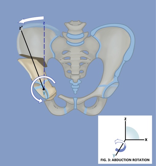
This results in greater and more three-dimensional reorientation of the acetabulum.
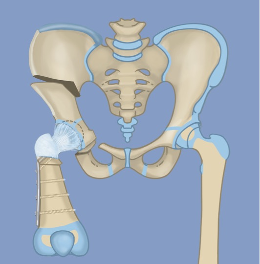
Next, the iliac bone crest is cut and the soft tissues reattached, as in the Dega osteotomy. The final step is to insert four screws into the pelvis in order to hold the alignment in place.
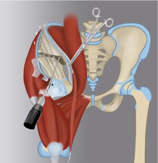
Ganz Osteotomy
In skeletally mature children and adults where there is no acetabular growth plate, a Ganz periacetabular osteotomy is used to reorient the acetabulum. In the Ganz osteotomy, five cuts to the pelvis are performed.
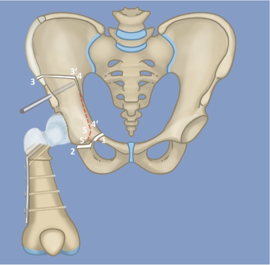
The released hip joint is then rotated twice in two different planes. It helps to visualize the pelvis along three different axes: the X, Y, and Z axes, as demonstrated below:
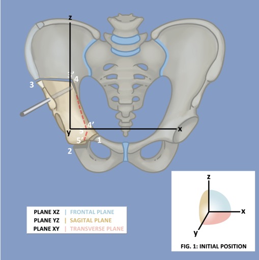
First, the pelvis is rotated in the Z-axis.

Second, the pelvis is rotated in the Y-axis.
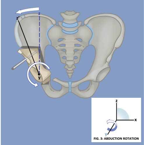
This results in the best three-dimensional reorientation of the acetabulum, however the growth plate is damaged in the process so this osteotomy is only viable in patients who have stopped growing.
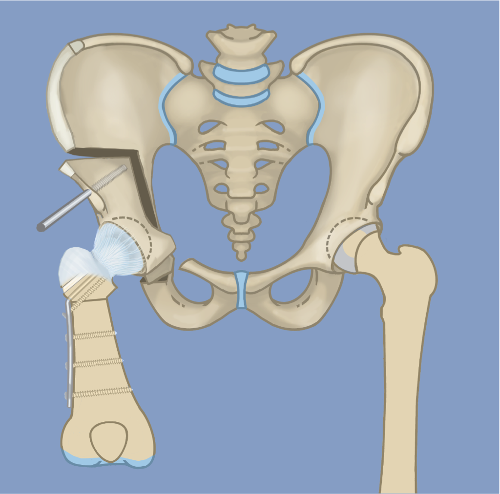
Next, the iliac bone crest is cut and the soft tissues reattached, as in the Dega osteotomy. The final step is to insert four screws into the pelvis in order to hold the alignment in place.

Type 1B CFD patients are distinguished from Type 1A by delayed ossification of the femoral neck. During the SUPERhip, BMP is inserted into the femoral neck and this will ossify into bone. This will essentially convert type 1B patients into type 1A. Once this occurs, type 1B patients are ready for lengthening. This process usually occurs within two years of the SUPERhip procedure, and lengthening is not performed until this conversion occurs.
To learn more about the SUPERhip procedure,
watch Dr. Paley's newest lecture.
Follow Up Care
After the SUPERhip (and possibly SUPERknee) surgery, the patient is placed in a spica cast for six weeks. Physical therapy will begin inpatient and aims to improve independence with bed mobility and transfers, and establish parent education. If deemed appropriate by Dr. Paley, the spica cast may be split so that it can be made into a removable cast. The cast is then removed for gentle daily physical therapy and to allow for easier hygiene and diaper change. Therapy involves passive flexion and extension of the hip and knee, not to exceed ninety degrees. Once the spica cast is removed, therapy will aim to gently maintain range-of-motion in order to prevent stiffness and speed recovery after the bone is healed and walking permitted, six weeks after surgery with a shoe lift. Most families will return home within a week or two after the superhip surgery and will receive therapy closer to home.
Long-term rehabilitation will include addressing residual muscle tightness and weakness with manual stretches, functional activities, and therapeutic exercises. There are typically no restrictions after twelve weeks, but it is important to gain clearance from Dr. Paley. The patient will be undergoing future lengthening and it is important to obtain full hip extension and prone (face-down) knee bending as preparation.